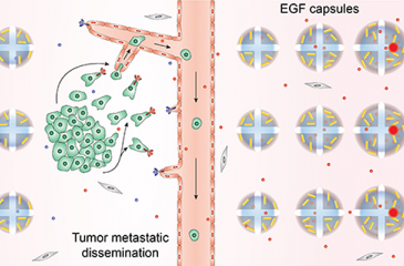
Researchers from both the University of Minnesota College of Science and Engineering and Medical School have created a new, dynamic 3D bioprinted tumor model that will help researchers screen anticancer drugs and study the spread of cancer.
The new model should help a current major pitfall of translating drugs from the lab bench to a viable treatment due to the differences of a 2D petri dish versus the 3D human body.
"I think our model has the key components to push the boundaries of in vitro cancer research,” said co-senior author Angela Panoskaltsis-Mortari, PhD, Department of Pediatrics.
The results of the study, published in Advanced Materials, found that the 3D-bioprinted model shows drugs take more time to kill fewer cells than previous studies have shown, however, the results likely show a more accurate representation.


